Dental Emergency
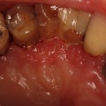
A dental emergency patient presented to my office with a non-healing ulcer located in the maxillary hard palate. This patient was 95 years old and had been treated with peridex antimicrobial mouth rinse for approximately 3 to 4 years. The ulcer was sensitive to palpate but did not cause any spontaneous pain. The patient was diagnosed with generalized chronic periodontitis, but I determined that it was a non contributory factor to the non-healing ulcer. I harvested a 1 centimeter biopsy of the ulcer and non affected tissue and sent it to OU pathology for histologic analysis.
Differential Diagnosis
The differential diagnosis includes
1. chronic fungal infection (Candidiasis)
2. Wegener’s Granulomatosis
3. Traumatic Ulcer
3. Squamous Cell Carcinoma.
Histologic Report
Invasive Squamous Cell Carcinoma
Background
Oral cancer accounts for less than 3% of all cancers in the United States, but it is the sixth most common cancer in males and twelfth most common in females. Approximately 94% of all oral malignancies are squamous cell carcinoma. Typically, the risk of intraoral cancer increases with age, especially for males. The cause of oral squamous cell carcinoma is multifactorial. No single causative agent or factor has been clearly defined or accepted, but both extrinsic and intrinsic factors may be at work. It is likely that more than a single factor is needed to produce such a malignancy. Extrinsic factors include such external agents as tobacco smoke, alcohol, syphilis, and sunlight. Intrinsic factors include systemic or generalized states, such as general malnutrition or iron-deficiency anemia. Many oral squamous cell carcinomas have been documented to be associated with or preceded by precancerous lesion, especially leukoplaia. Gingival and alveolar carcinomas are usually painless and most frequently arise from keratinized mucosa in a posterior mandibular site. They account for approximately 15% of all intraoral carcinomas.
Treatment
I immediately referred the patient to the OU Otolaryngologist department for treatment. Typically, intraoral squamous cell carcinoma treatment is guided by the clinical stage of the disease and consists of wide surgical excision, radiation therapy, or a combination of surgery and radiation therapy. The 5-year disease-free survival rate for intraoral carcinoma is 76% if metastasis has not occurred by the time of diagnosis, 41% when the cervical nodes are involved, and only 9% when metastasis below the clavicle is present.
Summary
Early diagnosis is key to patient treatment and survival. If the dentist is ever in doubt about a diagnosis (dental emergency), a biopsy should be completed or referred for a biopsy. Don’t wait!
-
- Dental Emergency, Non-Healing Ulcer


 by
by 